Arteriovenous Malformation (AVM) — Patient Guide
Find the Right Specialist — Free
Connect with top Arteriovenous Malformation specialists in Bangalore — 100% free, within 24 hours.
A Tangle of Blood Vessels That Can Bleed Without Warning
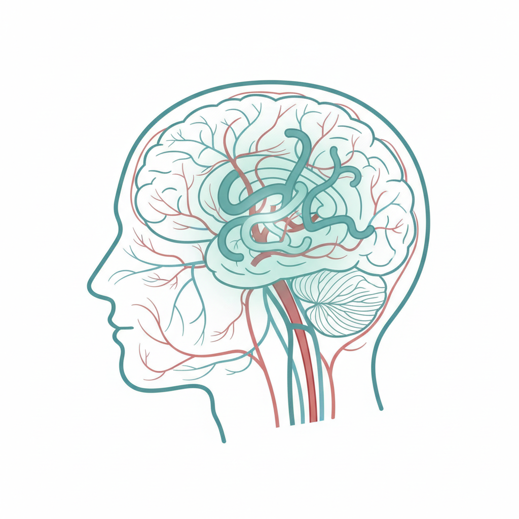
An arteriovenous malformation (AVM) is an abnormal tangle of blood vessels that connects arteries and veins in the brain, bypassing the normal capillary system. This short-circuit puts abnormal pressure on the vessels, which can cause them to rupture and bleed into the brain.
Most AVMs are present from birth and are discovered either after a bleed or incidentally during brain imaging. According to the Mayo Clinic, brain AVMs affect less than 1% of the population. With the right treatment, most patients can be treated.
What Is an Arteriovenous Malformation?
In a normal brain, arteries carry oxygenated blood to brain tissue through tiny capillaries before returning to the heart via veins. In an AVM, arteries connect directly to veins through a tangled mass of abnormal vessels called the nidus. This bypasses the capillaries, creating high-pressure flow that can damage surrounding brain tissue.
According to NCBI PubMed, approximately 50% of people with a brain AVM first present with a haemorrhage (bleed). The annual risk of bleeding from an untreated AVM is approximately 2 to 4% per year.
Symptoms of an AVM
Before bleeding: Headaches, seizures (most common presenting symptom), weakness or numbness in the limbs, vision problems, difficulty speaking.
After bleeding: Sudden severe headache, weakness or paralysis on one side, loss of consciousness, confusion, seizure.
A ruptured AVM is a medical emergency. Call 112 immediately.
Causes and Risk Factors
AVMs develop during foetal development. They are not inherited in most cases. Risk factors include male sex, rare family history, and hereditary haemorrhagic telangiectasia (HHT). Most AVMs occur without any identifiable cause.
How Is an AVM Diagnosed?
- MRI scan: First-line investigation showing the AVM location and relationship to brain structures.
- CT angiography (CTA): Used in emergencies to detect bleeding.
- Catheter angiography (DSA): Gold standard. Shows feeding arteries, nidus, and draining veins. Essential for treatment planning.
- Functional MRI: Maps the AVM relationship to critical brain areas before surgery.
Treatment Options
| Treatment | When It Is Used |
|---|---|
| Microsurgery | Small to medium AVMs in accessible locations. Immediate treat or manage if completely removed. |
| Stereotactic radiosurgery (Gamma Knife) | Small AVMs (under 3 cm) in deep areas. Takes 2 to 3 years to obliterate. |
| Endovascular embolisation | Used before surgery or radiosurgery to reduce blood flow. Rarely curative alone. |
| Active surveillance | Very small, asymptomatic AVMs in older patients. |
Surgery — What to Expect
Before surgery: Angiography review, embolisation 1 to 2 days before surgery, pre-operative blood tests and anaesthesia assessment.
During surgery: Craniotomy under general anaesthesia (4 to 8 hours). The neurosurgeon disconnects feeding arteries, removes the AVM nidus, and disconnects draining veins. Intraoperative angiography confirms complete removal.
After surgery: 1 to 2 days in ICU. Total hospital stay 5 to 10 days. Post-operative angiogram confirms complete AVM removal.
Recovery and Rehabilitation
- Week 1: Rest at home. Fatigue and mild headache are common.
- Weeks 2 to 6: Gradual return to light activities. Physiotherapy or speech therapy may begin.
- Months 2 to 3: Most patients return to desk work. Seizure medication continued for at least 1 year.
- Month 6+: Full recovery for most patients with small, unruptured AVMs.
Red flag symptoms: sudden severe headache, new weakness, confusion, seizure, or loss of consciousness — seek immediate care.
Cost of AVM Treatment in Bangalore
| Hospital Tier | Estimated Cost (INR) | What Is Included |
|---|---|---|
| Government / Trust Hospital | Rs. 2,00,000 to Rs. 4,00,000 | Basic microsurgery or embolisation, shared ICU |
| Mid-range Private Hospital | Rs. 4,00,000 to Rs. 7,00,000 | Semi-private room, intraoperative monitoring |
| Premium / Corporate Hospital | Rs. 7,00,000 to Rs. 15,00,000 | Private ICU, combined embolisation + surgery, Gamma Knife option |
Costs are estimates as of April 2026. Complex AVMs requiring multiple treatment sessions cost significantly more.
Insurance coverage: AVM treatment is covered under most private health insurance policies, CGHS, ESI, and Ayushman Bharat (PM-JAY) for eligible patients.
To get a personalised cost estimate from verified hospitals in Bangalore, submit your details on Patient-Help.com — free, confidential, within 24 hours.
How to Choose a Hospital in Bangalore
- NABH accreditation — national quality and safety standard.
- Cerebrovascular surgery volume — ask how many AVM cases the team performs per year.
- Biplane angiography suite — required for embolisation and intraoperative angiography.
- Gamma Knife availability — for patients who prefer radiosurgery.
- Multidisciplinary team — neurosurgeon, interventional neuroradiologist, and radiation oncologist.
Patient-Help.com matches you with verified specialists in Bangalore — free of charge.
Frequently Asked Questions
Is an AVM always dangerous?
An untreated AVM carries a 2 to 4% annual risk of bleeding. Over a lifetime, this risk accumulates significantly. However, small AVMs in non-eloquent brain areas may be monitored rather than treated, especially in older patients.
Can an AVM be treated?
Yes. Complete surgical removal or obliteration by radiosurgery treats the AVM and eliminates the risk of future bleeding. Surgery provides immediate treat or manage; radiosurgery takes 2 to 3 years to obliterate the AVM.
What is the Spetzler-Martin grade?
The Spetzler-Martin grading system (Grade I to V) classifies AVMs based on size, location in eloquent brain areas, and venous drainage pattern. Grade I and II AVMs are small and accessible — surgery is usually recommended.
How long does recovery take after AVM surgery?
For small, unruptured AVMs in accessible locations, most patients return to work within 4 to 8 weeks. Larger or ruptured AVMs may require 3 to 6 months of rehabilitation.
Is AVM treatment covered by insurance in India?
Yes. AVM treatment is covered under most private health insurance policies, CGHS, ESI, and Ayushman Bharat (PM-JAY) for eligible beneficiaries.
What happens if an AVM is left untreated?
An untreated AVM carries a cumulative risk of bleeding that increases over time. The consequences range from mild neurological deficits to severe disability or death. Most neurosurgeons recommend treatment for AVMs in younger patients.
Ready to find the right specialist for AVM treatment in Bangalore? Submit your details on Patient-Help.com and receive a free, confidential match with verified specialists — within 24 hours.
Medical Disclaimer: This article is for informational purposes only and does not constitute medical advice. Always consult a qualified specialist before making any healthcare decisions. Patient-Help.com connects patients with verified hospitals and doctors but does not provide medical diagnoses or treatment recommendations.
Sources
- Arteriovenous Malformation — Overview — Mayo Clinic
- Cerebral Arteriovenous Malformations — AANS
- Natural history and treatment outcomes of brain AVMs — NCBI PubMed
Find the Right Specialist — Free
Connect with top Arteriovenous Malformation specialists in Bangalore — 100% free, within 24 hours.
Find the Right Specialist — Free
Connect with top Arteriovenous Malformation specialists in Bangalore — 100% free, within 24 hours.
Medical Disclaimer: This article is for informational purposes only and does not constitute medical advice. Always consult a qualified healthcare professional for diagnosis, treatment, and medical decisions.